• Medical References

    Feel Free to download free e-book and powerpoint slides

  • Category name clash

    Lorem ipsum dolor sit amet, consectetuer adipiscing elit. Quisque sed felis. Aliquam sit amet felis. Mauris semper, velit semper laoreet dictum, quam diam dictum urna, nec placerat elit nisl in ...

  • Test with enclosures

    Here's an mp3 file that was uploaded as an attachment: Juan Manuel Fangio by Yue And here's a link to an external mp3 file: Acclimate by General Fuzz Both are CC licensed. Lorem ...

  • Block quotes

    Some block quote tests: Here's a one line quote. This part isn't quoted. Here's a much longer quote: Lorem ipsum dolor sit amet, consectetuer adipiscing elit. In dapibus. In pretium pede. Donec ...

Posted by medicalpitstop - - 0 коммент.

Urticaria or also known as hives


Def : hives are raised, often itchy, red welts on the surface of the skin. They are usually an allergic reaction to food or medicine


Causes:
allergic reaction to a substance- your body releases histamine and other chemicals into your bloodstream.- itching, swelling, and other symptoms.


When swelling or welts occur around the face, especially the lips and eyes, it is called angioedema. Swelling from angioedema can also occur around your hands, feet, and throat
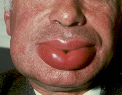
angioedema

Many substances can trigger hives, including:
  • Animal dander (especially cats)
  • Insect bites
  • Medications
  • Pollen
  • Shellfish, fish, nuts, eggs, milk, and other foods
Hives may also develop as a result of:
  • Emotional stress
  • Extreme cold or sun exposure
  • Excessive perspiration
  • Illness (including lupus, other autoimmune diseases, and leukemia,hepatitis B
  • Infections such as mononucleosis


Treatment

Treatment may not be needed if the hives are mild. They may disappear on their own. To reduce itching and swelling:
  • Avoid hot baths or showers.
  • Avoid irritating the area with tight-fitting clothing.
  • Take antihistamines. Diphenhydramine is considered the most effective.
If your reaction is severe, especially if the swelling involves your throat, you may require an emergency shot of epinephrine (adrenaline) or steroids. Hives in the throat can block your airway, making it difficult to breathe.


.
[ Read More ]

GN

Posted by medicalpitstop - - 0 коммент.


sign and symptoms
-hematuria(nephritic syndrome)
-protienuria(nephrotic syndrome)


type
-non-proliferative
-proliferative types
-primary(intrinsic)
-secondary(extrinsic)


what is the diff btwn non-proliferative and proliferative?


non-proliferative
Minimal change GN (also known as Minimal Change Disease)
Focal Segmental Glomerulosclerosis (FSGS)
Membranous glomerulonephritis


Thin BM nephropathy
etio-genetic causes
symptom-hematuria(rarely proteinuria)
diagnosis-biopsy electrom microscope(thinning of BM)
prognosis-benign(small risk to CRF) but can preceed to INC BP and proteinuria


Proliferative
IgA nephropathy (Berger's disease)
Post-infectious
Membranoproliferative/mesangiocapillary GN
Rapidly progressive glomerulonephritis


Minimal change GN (also known as Minimal Change Disease)
ages:children 80% while adult 20%
Etio-T-Lymphocytes mediated
Symptom-main-nephrotic syndrom,may also present with hematuria.
dignosis? selective proteinuria(albumin)


change: no change seen in light microscope only seen in electron microscope(accumulation of podocyte)
treatment:
1.supportive treatment- to decrease edema
2. steroid to halt the disease.(to those who are steroid resistance,use cyclophosphamide, cyclosporin(cytoxic theraphy)


*children more respond to theraphy
*3 months of treatment-deasea will cured


Focal Segmental Glomerulosclerosis (FSGS)
Syndrome : nephritic syndrome(nephotic syndrom according to oxford)
few focus of gemorules been fibrosis(sclerosis) with hyalinization of arterioles(IgM deposition0
maybe primary or secondary( reflux nephopathy,alport syndrome,heroin abuse)
treatment -corticosteroidand cyclosporine and cyclophosphamide (if streroid not effective)
Progressive until renal failure(30-50%)


Membranous glomerulonephritis
more frequent in adult(maybe heridetary,but only 9 cases in world)
what is it? thickening of glomerular basement membrane without hypercelular(diffuse granulat uptake of igG)
symptomes due to : hypersensitivity type 3-lead endothelial accumulation
symptoms? nephrotic(main) and nephritic symptoms
-high risk of vein thrombosis)
etiology : unknown ,maybe( cancer of lung or bowel,hepatitis or malaria drugs(penicillamine),autoimune dx(SLE)


how to diagnose? imunoflurescense
prognosis? Rule of third -one-third remain with MGN indefinitely, one-third remit, and one-third progress to end-stage renal failure


progress to? atrophy and hyalinisation(kidney keep to shrink)
treatment? corticosteroid


spike and dome explaination
These are due to membrane formation around the deposits, creating "spikes", followed by membrane formation over the deposits, creating the “domes”.   


proliferative type


Berger's disease( igA nephropathy)


age-young men have URTI (few attack)-overproduction of IgA


what is is?
deposition of IgA in mesingium lead to mesingium cell proliferation with increase matrix


what is IgA ?
antibody ussuakky present in mucus
have 2 type IgA and IgA1(ussually cause nephropatogenicity)


causes?
primary
secondary
-URTI
-Liver  disease
-GI infection
-henoch-schonlien purpura(what is it?vasculitis)
disease of the skin and other organs that most commonly affects children,causes palpable purpura (small hemorrhages); often with joint and abdominal pain.Withkidney involvement,


sign and symptomes
1.nephriti symptoms
-primaryly hematuria(see even gross)
-mild proteinuria (
2.occasinally nephrotic syndrom


pathogenesis?
plasma IgA increase ,IgA circulating contain imune complex.deposition of imune complex lead nephropathy


TX immunosuppresion drug(cyclophosphamide and mycophenolate)


20% of cases lead to ESRF worse if the is INC BP,male and have proteinuria


Post-Infectious
etiology- any infections espcially streptococus pyogenous
time to occur- 10-14 days after infection(pharyngitis or skinn lesion)


sign n symptome _glomerularnephritis(nephritic(main) and nephrotic symptoms) 


diagnosis
1.history of disease(anamnesis)
2.streptococus titer(antistreptolysin O titers)


microscopic changes
-hypercellurarity of messingial cell and endothelial in capaliry
lead to influx of neutrophil and monocytes in urine
(in some situation hypercelularity of mesingial and endocapaliry lead to cpmpression of capsule bowman(cresentic formation)


progression?
ussually 2-4 weks resolve without complication


membranoproliferative?mesinggiocapalliry GN
most severe,why?
rapid progression to ESRF over weeks(steroid maybe help to halt the progression)


main presentaion -nephrotic syndrome


causes?
2 main disease lead to RPGN
1.Goodpasture syndrome?
-autoimune dx against BM of kidney and lungs.
-s n s-hemoptysis
-histologically -we can see deposition of IgG
-tx-imunosupressive therapy( steroid-methylprednisolone) and plasmapheresis(extraction of plasma remain the RBC)


2. vasculitic dx
-wegener dx(vaslulitis in organ like kidney and lung)
-blood test-ANCA not imune complex deposition


histologically(general)
-cresent(why? fibrin in capsule bowman-increase permeability-stimulate proliferation of parietal cell and influx of monocytes)
rapid growth of fibrosis lead to compression of capallary loops and decrease capsule bowman space


how to examine the patient?
1.dx history
-diabetes m
-hypertension
-sore throat(post infectious GN)
-medication (nsaid)
-family hx(sickel cell dx)
-neoplasm


2.general examination
-edema(face,pitting edema of the leg or hand)
-ridge of the nail(sign of hypoproteinimia)
-pallor(?)


ridge of the nail


what to check?
1.ascitis
2.protein n sugar in urine
3.24 hours urine protein ,creatinine clearence and albumin
4.FBC(full blood count) , ESR and crp
5.serum cholesterole
6.DNA antibody,ANCA,ANA,anti-GBM
7.renal ultrasound andrenal biopsy


how to manage?
1.refer t nephrologist
-by keep the BP less than 130/80 or less125/75 if proteinuria more than 1g/d


what is nephtrotic syndrome?
-albuminuria
-hypoalbuminimia
-edema
-lipiduria
-hyperlipidemia(hyperfunction of liver) why?


complication?
1.thoboembolic event(loss of antitrombin 3 factor..which produce in liver and has bee loss trough kidney)
2.protein malnutrition
3.athereosclerosis(hihg lipid production)
4. infection


What is pitting edema?
Pitting edema can be demonstrated by applying pressure to the swollen area by depressing the skin with a finger. If the pressing causes an indentation that persists for some time after the release of the pressure


CAuses of non pitting edema..
Non-pitting edema can occur in certain disorders of the lymphatic system such as lymphedema, which is a disturbance of the lymphatic circulation that may occur after a mastectomy, lymph node surgery, or congenitally. 


What is nEPHROTIC SYNDRME ACTUALLY?
1.proteinuria more than 3g/24h
2.hypoalbumininia (less 30g/L)
3.edema


what will happen?
loss protein-decrese albumin-decrease oncotic pressure in blood vessel-fluid go to extracellular space-plasma oncotic pressure remain the same-lead to sodium retention-edema


diff diagnose?
cardic failure(pulmonary edema,mild proteinuria)
liver dx (decrease in albumin level)


complication?
1.inc susceptibility to infection-Ig leak to urine
2.thromboembolismm-inc clotting factor(hyperfx of liver)
3.hyperlipidemia-(the same reason-in respond to dec oncotic pressure)


what we have to do?
1.if the is ni family history or other previous dx-we gave them steroid (why? most of the case due to minimally change GN whic can be cure by steroid therapy)
2.but if the protenuria  is not regress in 1 months ..we have to do renal biopsy)


Tx
1.restrict salt intake
2.diuretic (furasemid
3.infection?(give vaccination)
4.prophylactic (heparin)
5.hypertension(ace i and A2A)


[ Read More ]

Posted by medicalpitstop - - 0 коммент.

5 part of trauma
-head(most severe cases need many types of doctor)
-neck
-extremity(most frequent cases)
-abdomen
-pelvic

center of emergency in moscow
-metro сухаревькая

type of trauma
-open trauma (10%)
-close trauma (90%)

classification of chest trauma
-internal organ involvement
1.with ribs involvement(53%)
2.w/o ribs involvement (47%)

-no internal organ involvement
1.w ribs involment(19%)
2.w/o ribs involvement (8%)

what is flaily chest?

Complication?
1.pneomothorax
2.hemothorax
3.pneumohemathorax
4.lesion of organ
-liver
-spleen
-kidney
-diaphragm

organ trauma frequency
1.lung 90%
2.heart 8%
3.diaphragm 4%
4.large vessel 3%
5.bronchus 1.5%
6.trachea 1%

symptoms
1.pneumathorax(can make diagnoais by palpation-lienek's idea)
2.emphysema
3.hemothorx
4.pneumothorx

how to examine the patient?
1.position of the patient
2.color skin on trauma area
3.deformation
4.crepitation
5.skin lesion

how to make diagnosis?
1.x-ray(gold standard
2.puncture(when there is fluid )
3.meningealscopy,thoncoscopy
4.CT and MRI

acute lung lesion
-direct
yshib
internal hematoma
razrez
segmet,lobe

-mix

-indirect
fat emboli(dangerous-can go to brain lead to stroke)
repiratoey distress syndrom
edema(inrstitial alveolar)

what is index oxygenation?
Pa02/fi02
arterial oxygen /pumonary oxygen

if fi02 is more than 60%

How to treat?
1.stabilization of chest tissue
2.ventilatory help
3.nitrogen oxygen-vasodilator

what is polyporphine?
Synthetic polypoephine


2.emphysema
[ Read More ]

Posted by medicalpitstop - - 0 коммент.


>50% of significant chest trauma cases due to blunt trauma
Compressional forces flex and fracture ribs at weakest points
Ribs 1-3 requires great force to fracture
  •      Possible underlying lung injury
Ribs 4-9 are most commonly fractured
Ribs 9-12 less likely to be fractured
  •      Transmit energy of trauma to internal organs
  •      If fractured, suspect liver and spleen injury
Hypoventilation is COMMON due to PAIN


Rib Fractures with Open Pneumothorax


QUIZ : 
  • What Is Open Pneumothorax? 
  • How does it differ to "Closed Pneumothorax" and "Tension Pneumothorax"?
  •  What is the managements for Pneumothorax patients?


Management of Open Pneumothorax

  • Cover site with sterile occlusive dressing taped on three sides

[ Read More ]

Posted by medicalpitstop - - 0 коммент.

Pathophysiology of Thoracic Тrauma


uBlunt Trauma
Results from kinetic energy forces
Subdivision Mechanisms
uBlast
Pressure wave causes tissue disruption
Tear blood vessels & disrupt alveolar tissue
Disruption of tracheobronchial tree
Traumatic diaphragm rupture
uCrush (Compression)
Body is compressed between an object and a hard surface
Direct injury of chest wall and internal structures
uDeceleration
Body in motion strikes a fixed object
Blunt trauma to chest wall
Internal structures continue in motion
uLigamentum Arteriosum shears aorta
Age Factors
uPediatric Thorax:  More cartilage = Absorbs forces
uGeriatric Thorax:  Calcification & osteoporosis = More fractures


uPenetrating Trauma
Low Energy
uArrows, knives, handguns
uInjury caused by direct contact and cavitation
High Energy
uMilitary, hunting rifles & high powered hand guns
uExtensive injury due to high pressure cavitation

Shotgun
uInjury severity based upon the distance between the victim and shotgun & caliber of shot
uType I:  >7 meters from the weapon
               –Soft tissue injury
uType II:  3-7 meters from weapon
               –Penetration into deep fascia and some internal organs
uType III:  <3 meters from weapon 
               –Massive tissue destruction


Injuries Associated with Penetrating Thoracic Trauma


uClosed pneumothorax
uOpen pneumothorax (including sucking chest wound)
uTension pneumothorax
uPneumomediastinum
uHemothorax
uHemopneumothorax
uLaceration of vascular structures
uTracheobronchial tree lacerations
uEsophageal lacerations
uPenetrating cardiac injuries
uPericardial tamponade
uSpinal cord injuries
uDiaphragm trauma
uIntra-abdominal penetration with associated organ injury


[ Read More ]